Figure 5.
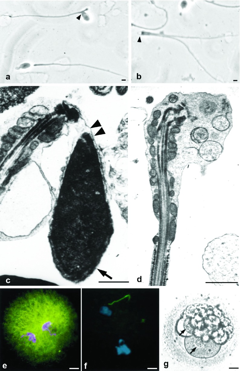
Pathologies of the sperm neck. (a, b) Sperm smear showing abnormal alignment of heads and tails (a, arrowhead) and acephalic spermatozoa (b, arrowhead). (c) Electron microscopy shows lack of attachment between head and mid-piece (double arrowhead) and normal acrosome (arrow). (d) Acephalic spermatozoon: absence of the head. The enlarged cephalic end is occupied by the mitochondrial sheath, connecting piece and vesicular structures. (e, f) Heterologous ICSI. (e) Normal pronuclear and aster formation (microtubules, green fluorescence) after injection of a normal human spermatozoon into a bovine oocyte. (f) When spermatozoa with abnormal head–mid-piece relationships are injected pronuclei form, but there is no aster development in the zygote. (g) Human ICSI failure after injection of spermatozoon with neck anomalies. Pronuclei are visible (arrows), but syngamy and cleavage did not occur and the zygote degenerated. Bars=1 µm (a–d) and 20 µm (e–g). g is reproduced with permission from Chemes et al.49 e and f are reproduced with permission from Rawe et al.69

