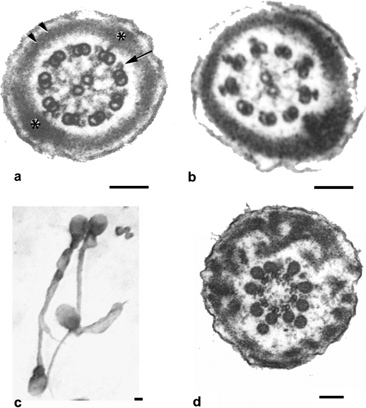
Figure 6.
Flagellar pathologies. (a) Transverse section of a normal sperm principal piece. The classical 9+2 structure of the axoneme is clearly seen. Note dynein arms (arrow) and normal fibrous sheath (asterisks and double arrow). (b) Lack of dynein arms in microtubular doublets of immotile spermatozoon in a patient with primary ciliary dyskinesia. (c, d) Dysplasia of the fibrous sheath. Tails are short, thick and of irregular profile (c). Ultrastructural examination depicts thickened fibrous sheath and the absence of the central microtubules of the axoneme. Bars=0.1 µm (a, b and d) and 0.5 µm (c). Flagellar diameter is 0.4 µm (a and b) and 0.7 µm (d). a is reproduced with permission from Turner et al.93 Figure 6d is reproduced with permission from Chemes et al.9

