
FIG 4 .
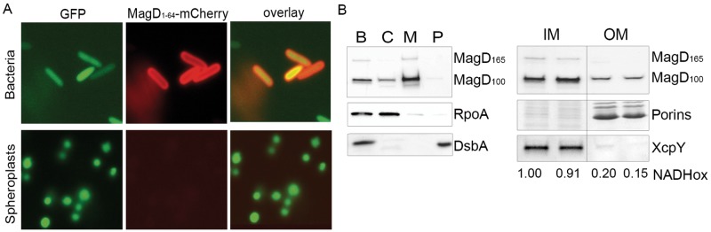
MagD is translocated to the periplasm but appears associated with the inner membrane. (A) The gene encoding MagD1-64-mCherry was expressed under the arabinose-inducible promoter carried within pJN105. The plasmid was introduced into different P. aeruginosa strains expressing cytoplasmic GFP. Spheroplasts obtained by lysozyme treatment and bacteria were observed by fluorescence microscopy using appropriate filters. Note the peripheral labeling in bacteria and the absence of labeling in spheroplasts. (B) Fractionation of PAO1 and immunoblot analyses of MagD. Whole bacterial cells (lane B) were fractionated into the cytoplasm (lane C), total membranes (lane M), and periplasm (lane P) and immunoblotted with anti-MagD antibodies. RpoA and DsbA were used as cytoplasmic and periplasmic markers, respectively. Total membranes were further separated by centrifugation on a sucrose gradient. Two inner membrane (IM) and two outer membrane (OM) fractions were analyzed. The measurement of NADH oxidase activity (NADHox) and immunoblotting with anti-XcpY antibodies were used as IM markers. The OM is characterized by porins (36 kDa) visualized by Coomassie blue staining. MagD is preferentially present in the IM.