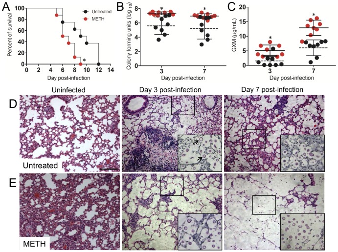
FIG 1 .
METH exacerbates cryptococcosis. (A) Survival differences of METH- and PBS-injected (untreated, control) C57BL/6 mice after intratracheal (i.t.) infection with 107 C. neoformans cells (n = 5 per group). Asterisks denote P value significance (P < 0.01) calculated by log rank (Mantel-Cox) analysis. This experiment was performed twice, and similar results were obtained. (B) Lung fungal burden (numbers of CFU) in untreated and METH-treated mice infected with 106 C. neoformans yeast cells. (C) C. neoformans GXM released in lung tissue. (B and C) Each circle represents the value for 1 mouse (n = 8 per group). Solid and dashed lines represent average results for METH-treated and untreated groups, respectively. Error bars denote standard deviations. Asterisks denote P value significance (*, P < 0.05) calculated by Student’s t test at each time point. These experiments were performed twice with similar results. (D and E) Histological analysis of lungs removed from untreated (D) and METH-injected (E) C57BL/6 mice. Representative H&E-stained sections of the lungs are shown. The large insets are magnifications of the smaller boxes in the H&E-stained sections to better show the cryptococcal cells stained with mucin carmine (indicated with arrows in the middle image of panel D; 3 days postinfection; untreated). Bars, 10 µm.

