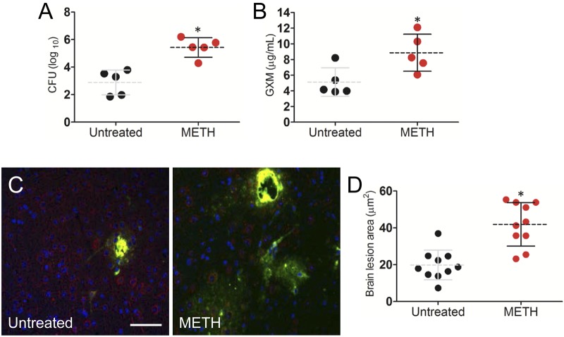
FIG 6 .
METH significantly induces C. neoformans capsular polysaccharide release in the CNS 3 days after pulmonary infection. (A) The brain fungal burdens in METH-treated mice infected intratracheally with 106 C. neoformans cells were significantly greater than those in untreated mice (n = 5 per group). (B) Capsular polysaccharide concentrations in the supernatants of C. neoformans-infected brain homogenates of untreated or METH-treated mice (n = 5 per group). (A and B) Dashed lines represent the mean values for the METH-treated and untreated groups; error bars denote standard deviations. Each circle represents the value for 1 mouse. P values (*, P < 0.05) were calculated by Student’s t test. (C) Immunofluorescence staining of brain lesions (cryptococcomas) caused by C. neoformans in untreated or METH-treated animals. Capsular-polysaccharide-specific MAb 18B7 (yellow) was used to label fungal cells. MAP-2 (red) and DAPI (blue) staining were used to label the cell bodies and nuclei of neurons, respectively. Bar, 5 µm. (D) Area analyses of brain lesions caused by C. neoformans strain H99 cells were performed in tissue sections of untreated or METH-treated mice (n = 3 per group). The areas of 10 brain lesions per condition were measured using ImageJ software. Each circle represents a single lesion. Dashed lines and error bars denote averages of 10 measurements and standard deviations, respectively. Asterisks denote P value significance (*, P < 0.001) calculated by the t test. (A to D) The experiments were performed twice, with similar results.

