Fig. 2.
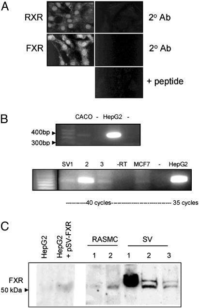
FXR is expressed in vascular smooth muscle cells. (A) Immunofluorescence for FXR and RXR in RASMC. The 2°Ab is negative control for when primary antibody was omitted; + peptide is immunofloresence of FXR, performed with primary antibody preabsorbed with blocking peptide. (B) RT-PCR for hFXR. (Upper) FXR as a band at 362 bp in the positive controls CACO and HepG2; -, RT-PCR when reverse transcriptase was omitted. (Lower) RT-PCR for hFXR on hSVSMC (SV1-3) from three patients, -RT where reverse transcriptase was omitted (for SV3), MCF-7 cells (-RT), and in the positive control HepG2 cells. (C) Western blot analysis for FXR in HepG2 cell (transfected empty vector), HepG2 cells transfected with rat FXR (24 h), RASMC (1, WKY3m-22; 2, primary RASMC), and hSVSMC (SV1-3 from three patients). Note that SV sources 1-3 from RT-PCR do not correspond to SV 1-3 in Western blot.

