
Figure 4.
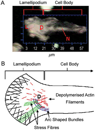
(A): Optical image of a motile prostate cancer (PCa) cell with a lamellipodial extension (L) projecting out in the direction of cellular travel. N, Nucleus; D, de-polymerized actin filaments. (B): Schematic showing the internal architecture of the lamellipodium (L), with actin filaments linking the internal cellular structure to the external lipid cellular membrane. These filaments undergo a constant process of polymerization and destruction (D), resulting in movement of the lamellipodium and forward movement of the cell (Reproduced with permission from2).