
Figure 8.
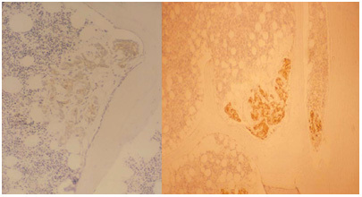
Photomicrographs of two bone biopsies taken from different prostate cancer (PCa) patients. These show the typical appearance of early micro-metastases from PCa. The epithelial cells are stained for prostate-specific angiten (PSA) and appear brown in this image. The cell colonies lie close to the bone surface (uniformly pale), where they stimulate early osteoblastic activity. In the surrounding bone marrow, the fibroblasts are stimulated to induce a desmoplastic reaction. There is no evidence of bone resorption at this stage of the disease process (Reproduced with permission from2)