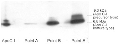
Figure 4.

ApoC-I Western blotting of the serum fractions by HPLC purification. A representative case (case 1) of Western blotting of patient serum that shows two bands of ApoC-I (a 9.3-kDa precursor and a 6.6-kDa mature protein). The intensity of these two ApoC-I bands increased as the disease progressed, similar to what was observed with the SELDI-TOF-MS analysis (shown at points A, B and E).
