
Figure 2.
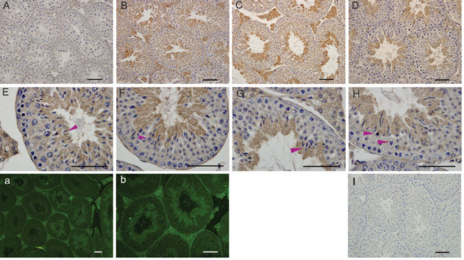
Immunolocalization of ubiquitin-specific protease 26 (USP26) protein in the developing mouse testis. USP26-positive cells are characterized by brown staining as a result of the DAB colorimetric reaction in (A–H) and in green as a result of FITC immunofluorescence staining in (a) and (b). USP26 immunostaining is absent in 20 day (A), weak in 30 day (B) and intense in 35 and 45-day mouse testes, in which the staining is strong in the cytoplasm of condensing spermatids (steps 9–16) (C and D). (E-G) show seminiferous epithelium at late(E) and early stages(F, G) respectively, and the arrowheads in (H) point to cells undergoing meiotic metaphase. Immunofluorescence staining in panels (a) and (b) also shows USP26 expression in spermatids and in Leydig cells in testes. I: Control section. Scale bars = 50μm.