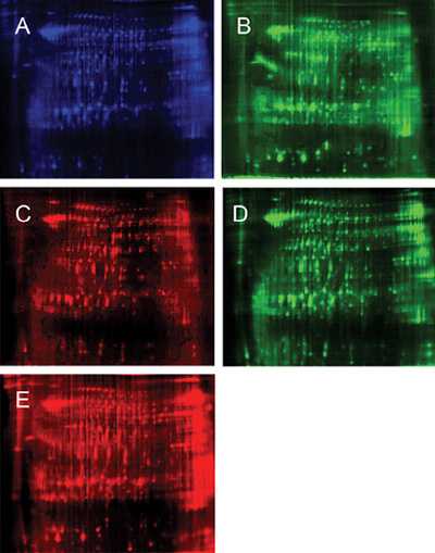
Figure 1.

Schematic description of the mixing of separately labelled samples onto the same gel, with subsequent independent Cy-dye imaging for data analysis. The representative two-dimensional (2-D) difference gel electrophoresis (DIGE) image of the two pools (24 cm, PH3-10, normal limits [NL]). Protein extracts (50 mg each) were covalently conjugated to two different fluorescent dyes, and the extracts were pooled and separated on the same gel. The first (horizontal) dimension was a PH 3–10 non-linear focusing gradient and the second vertical dimension was a 12.5% homogenous SDS polyacrylamide gel. Cy2 (blue) was conjugated to proteins used as an internal standard in all samples. In one gel, Cy3 (green) was conjugated to proteins from the round-headed sample (A) and Cy5 (red) was conjugated to proteins from the normal sample (B). In another gel, proteins from the normal sperm sample were conjugated to Cy3 (C) and proteins from the round-headed sperm sample were conjugated to Cy5 (D). (A) Mixed sample labelled with Cy2; (D) Round-headed sperm labelled with Cy3 in gel 1; (c) Normal sperm labelled with Cy5 in gel 1; (D) Normal sperm labelled with Cy3 in gel 2; (E) Round-headed sperm labelled with Cy5 in gel 2.
