
Figure 2. Among PC Synthesis Enzymes, CCT Uniquely Localizes to LDs.
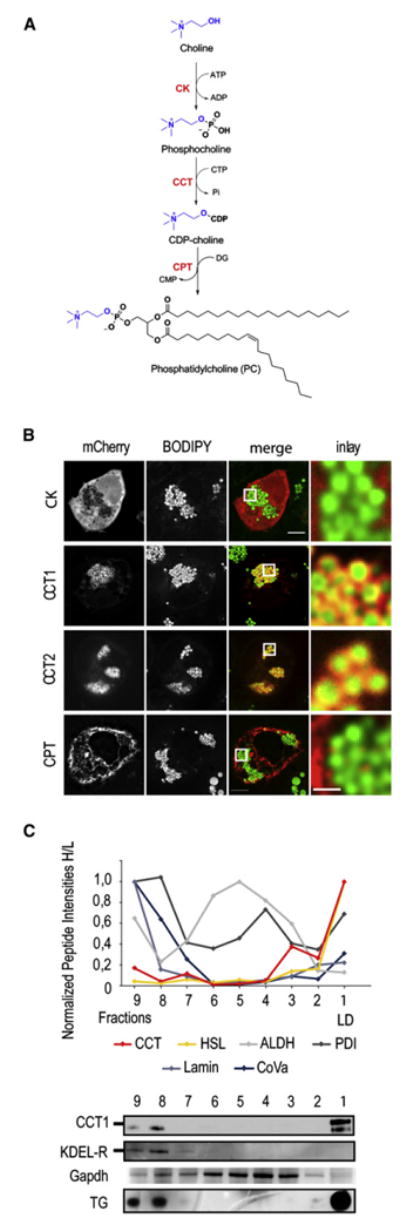
(A) Overview of the reactions of the Kennedy pathway.
(B) Transiently expressed mCherry-tagged CCT1 and CCT2, but not CK and CPT, (left panels) localize to LDs in S2 cells loaded with 1 mM oleate for 12 hr. Overlays and a magnification are shown (right two panels). Bar, 5 μm (overview) or 1μm (magnification).
(C) Endogenous CCT localizes to LDs. Normalized peptide intensities determined by LC-MS/MS are shown for fractions of a LD purification. CCT, hormone-sensitive lipase (HSL, LD marker), disulfide isomerase (PDI; ER marker), lamin (nuclear marker), cytochrome oxidase subunit Va (CoVa; mitochondrial marker), and aldehyde dehydrogenase (ALDH; cytosol marker) are shown. Bottom panels show western blots for CCT1, KDEL receptor, and Gapdh and a TLC for TG of each fraction normalized to protein.