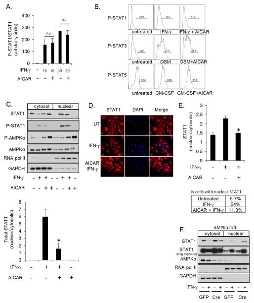
Figure 5. AMPK Signaling Does Not Influence STAT1 Phosphorylation but Attenuates Nuclear Translocation.
A. Astrocytes were stimulated with IFN-γ (10 ng/ml, 30 min) in the absence or presence of AICAR (1 mM) followed by quantification of immunoblots for P-STAT1 and STAT1. Values are expressed as the ratio of P-STAT1 to total STAT1. B. Astrocytes were stimulated with IFN-γ (10 ng/ml, 30 min), OSM (1 ng/ml, 30 min) or GM-CSF (10 ng/ml, 30 min) in the absence or presence of AICAR (1 mM) and P-STAT1, P-STAT3 and P-STAT5 measured by flow cytometry. C. Astrocytes were stimulated with IFN-γ (10 ng/ml) for 30 min in the absence or presence of AICAR (1 mM) followed by fractionation into cytosolic and nuclear fractions. STAT1, P-STAT1, AMPKα and P-AMPK subcellular distribution was examined by immunoblotting. The results for total STAT1 were quantified. The separation of GAPDH and RNA pol II demonstrates efficient separation of cytosolic and nuclear fractions, respectively. D and E. STAT1 localization was examined by immunofluorescent microscopy and quantified. The percentage of cells with nuclear/cytosolic STAT1 > 2 is reported in the table. F. AMPKαfl/fl astrocytes were transduced with GFP or GFP-Cre adenovirus followed by stimulation with IFN-γ (10 ng/ml, 30 min) and subcellular fractionation. N=3, *p < 0.05.

