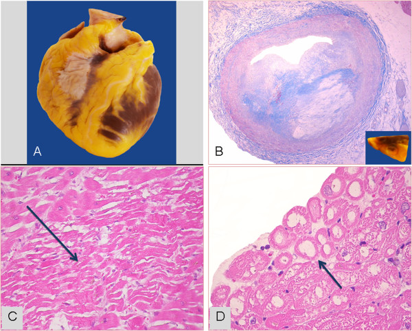
Figure 3.
The donor heart unsuitable for transplant of Figure 2. A) Macroscopic aspect of donor heart. B) Fibro-lipidic plaque narrowing (≈90%) the lumen of left anterior coronary artery (Mallory trichrome stain; original magnification 25x); in the inset, the corresponding macroscopic sample. C) Focus of myocardial coagulative necrosis (arrow) in the left ventricle (Hematoxylin-Eosin stain; original magnification 200x). D) Subendocardial coagulative myocytolysis (arrow) in the septal myocardium (Hematoxylin-Eosin stain; original magnification: 400x).

