Figure 4.
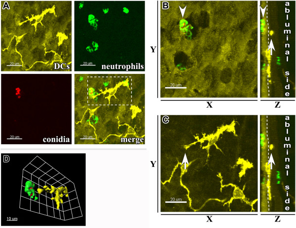
Internalization of A. fumigatus conidia in the epithelial compartment of conducting airways. (A) Representative confocal image taken from the proximal ventral segment of the whole-mount airway main axial pathway showing the distribution of MHC II+ epithelial DCs (upper left panel: MHC II+ DCs, yellow; epithelium auto-fluorescence, dark yellow), Ly-6G+ neutrophils (upper right panel, green), A. fumigatus conidia (lower left panel, red), and a merged image (lower right panel) at 4 hours post-conidial application. Scale bar = 20 μm. (B, C) The neutrophil locating in close proximity to the luminal side of the epithelium (arrowhead) and the intraepithelial DC (arrow) are indicated on Z-projection (left panels) or on X-projection (right panels) of the area displayed in (A). (D) The same neutrophil and DC are displayed on the three-dimensional larger magnification image of the region boxed in (A). Scale bar = 20 μm (A, B, C); and 10 μm (D).

