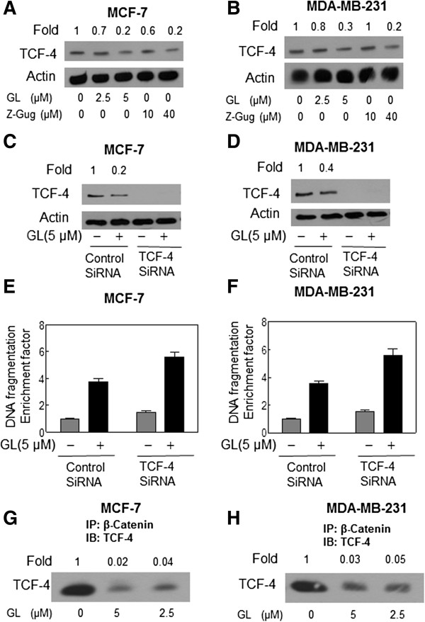
Figure 6.

Immunoblotting for TCF-4 using lysates from MCF-7 (A) or MDA-MB-231 (B) cells treated with DMSO (control) or 5 μmol/L GL for 24 h. Immunoblotting for TCF-4 using lysates from MCF-7 (C) or MDA-MB-231 (D) cells transiently transfected with a control nonspecific siRNA or TCF-4 -targeted siRNA and treated for 24 h with DMSO or 5 μmol/L GL. The blots were stripped and reprobed with anti-actin antibody to ensure equal protein loading. The numbers on top of the immunoreactive bands represent changes in protein levels relative to DMSO-treated control cells (A-B) or relative to DMSO-treated nonspecific control siRNA–transfected cells (C-D). Cytoplasmic histone-associated apoptotic DNA fragmentation in MCF-7 (E) or MDA-MB-231 (F) cells transiently transfected with a control nonspecific siRNA or TCF-4-targeted siRNA and treated for 24 h with DMSO or 5 μmol/L GL. The results are expressed as enrichment factor relative to DMSO-treated control cells transiently transfected with the control nonspecific siRNA. Each experiment was done twice, and representative data from a single experiment are shown. Columns, mean (n=3); bars, SE. *, P<0.05, significantly different between the indicated groups by paired t test. GL treatment inhibited β-Catenin binding to TCF in human breast cancer cells. Immunoblotting for TCF-4 using β-Catenin immunoprecipitates from MCF-7 (H) and MDA-MB-231 (G) cells treated for 24 h with DMSO (control) or GL 2.5 and 5 μM. The numbers on top of the immunoreactive bands represent change in levels relative to DMSO-treated control for each cell line. Each experiment was performed at least twice using independently prepared lysates.
