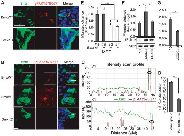
Fig. 5. BMX regulates FAK-mediated migration.
(A) 293 cells transfected with BmxWT or BmxKD were immunostained for BMX and pTyr576/Y577 FAK. Scale bars, 20 μm. Images are representative fields from one of three independent experiments. (B) Scratch wounds were introduced into COS7 cells transfected and immunostained as in (A), and pictures of the leading edge (arrows) were taken after 3 hours. Scale bars, 20 μm. Boxed region is magnified in the middle row. Images are representative fields from one of three independent experiments. (C) BMX (green) and pTyr576/Y577 FAK (red) fluorescence intensities along the axis defined by the white arrows in (B) were assessed using Zeiss LSM (WT, above; KD, below). Circles denote the leading edge. Data are results from scanning 10 cells each transfected with BmxWT or BmxKD. (D) Colocalization of BMX and pTyr576/Y577 FAK in lamellipodia at leading edges (n = 22) versus random areas (n = 20) from three independent experiments was assessed using Volocity. (E) Scratch wounds were introduced into confluent Bmx+/−, Bmx+, or Bmx− MEFs, and leading edges were photographed at 0 and 9 hours. Migrated distance was normalized to the #1 Bmx− group. Data are means ± SEM from three experiments. ***P < 0.001. (F) Scratch wounds were introduced into confluent Bmx− MEFs stably overexpressing BmxWT, BmxKD, or empty vector (EV) and photographed as in (E). Migrated distance was normalized to the EV group. Data are means ± SEM from three independent experiments. *P < 0.05. (G) Bmx− MEF cells stably overexpressing BmxWT were preincubated with FAK inhibitor (PF562271) for 4 hours. Scratch wounds were introduced and photographed as in (E). Migrated distance was normalized to the control group. Data are means ± SEM from three experiments. ***P < 0.001.

