
Figure 2.
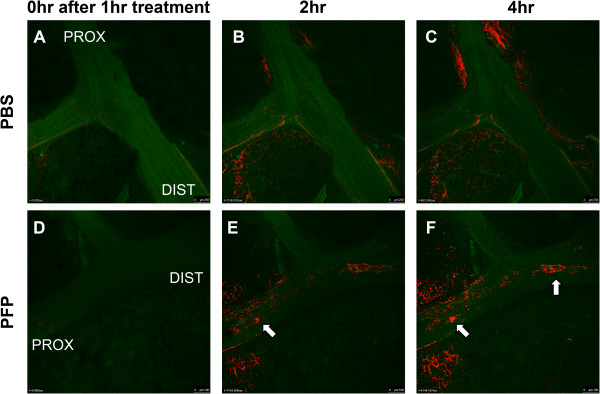
Ex vivo oxidative stress detection. Rat right cranial lobes were dissected to expose the main axial airway and airways were treated with either PFP or PBS. Lungs were incubated with CellROX, a fluorescent dye that indicates oxidative stress, and washed with Live Cell Imaging solution after 1 hour of exposure and imaged continuously. Representative pictures from lungs taken directly (A, D), 2 hours (B, and E) and 4 hours (C and F) after a 1 hour treatment of either PBS (A-C) or PFP (D-F). Substantially more CellROX fluorescence could be observed in the airway lumen of the PFP-treated lung compared to PBS controls in a time dependent and proximal (PROX) to distal (DIST) manner. Focal patches of cells with oxidative stress were observed (arrows) increased over time in the PFP-treated lungs.