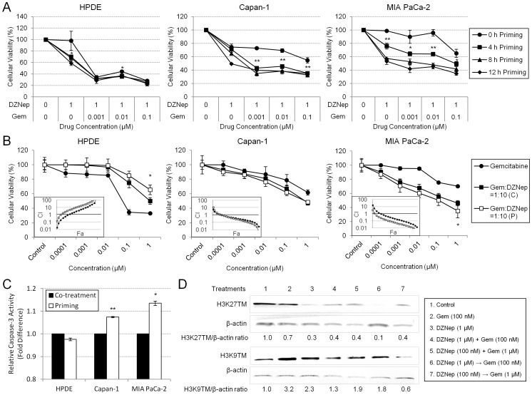
Figure 5. Short priming of DZNep demonstrated superior cytotoxicity and synergy with gemcitabine than co-exposure of the two drugs.
A. Short exposure with DZNep for 4–8 h produced maximal cytotoxic effects. Cells were exposed with DZNep at 1 µM for varying time intervals followed by increasing concentrations of gemcitabine (0–0.1 µM). Significance between 0 and 4 h is indicated. B. Superior cytotoxicity and synergism between gemcitabine and DZNep were observed when cells were primed with DZNep, as opposed to cotreatment with gemcitabine. Representative growth inhibition curves are shown. Twenty-four hours after 3×10³ cells/well were seeded in a 96-well plate, cells were exposed to gemcitabine and DZNep concentrations at a 1∶10 ratio either as a co-treatment for 72 h (C) or a primed treatment (with DZNep for 8 h followed by gemcitabine for 72 h) (P). Cellular viabilities were measured using MTT assays. Significance between co-treatment and priming is indicated. Combination index (CI) plots (insets) show the interactions between the two drugs. CI>1, antagonism; CI = 1, additivity; CI<1, synergism. Bars, SD. n = 3. *p<0.05, **p<0.01. C. Apoptosis levels were significantly greater in Capan-1 and MIA PaCa-2 cells with priming compared with co-treatment, while apoptosis levels in HPDE decreased with priming. Cells were either co-treated with 10 µM DZNep and 1 µM gemcitabine or primed with 10 µM DZNep for 8 h followed by 1 µM gemcitabine. Fluorescence values were background-subtracted and are indicated as fold-change from co-treatment to priming. Significant differences between co-treatment and priming were identified using the Student’s t test. Bars, SD. n = 3. *p<0.05, **p<0.01. D. Maximal reduction in H3K27 trimethylation was seen with priming schedules at 1∶10 DZNep:gemcitabine. MIA PaCa-2 was treated with vehicle, gemcitabine for 72 h, DZNep for 8 h, DZNep and gemcitabine for 72 h, or DZNep for 8 h followed by gemcitabine for 72 h. 100 µg of whole cell lysates were subjected to Western blotting analysis. Blots were stripped and re-probed for β-actin, the internal loading control. Densitometry ratios are indicated.

