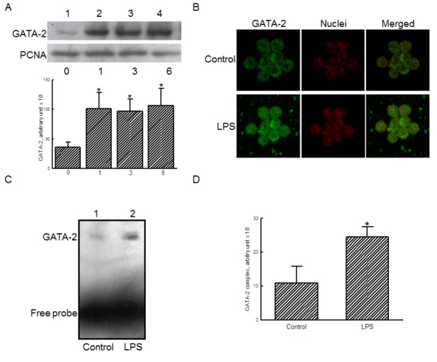
Figure 2. Effects of lipopolysaccharide (LPS) on the translocation and transactivation of GATA-2.

RAW 264.7 cells were exposed to 100 ng/ml LPS for 1, 3, and 6 h. Amounts of nuclear GATA-2 were immunodetected (A, top panel). Levels of nuclear PCNA were measured as the internal control. These immunorelated proteins were quantified and statistically analyzed (bottom panel). RAW 264.7 cells were treated with 100 ng/ml LPS, the translocation of GATA-2 from the cytoplasm to nuclei were analyzed using confocal microscopy (B). The transactivation activity of GATA-2 was assayed using an EMSA analysis (C). These DNA-protein bands were quantified and statistically analyzed (D). The immunoblotting, confocal, and DNA-protein binding results shown are a representative of at least 3 experiments, and the other statistically analyzed results are a compilation of 6 replications. Each value represents the mean ± SD. An asterisk (*) indicates that the value significantly differed from the respective control, p < 0.05.
