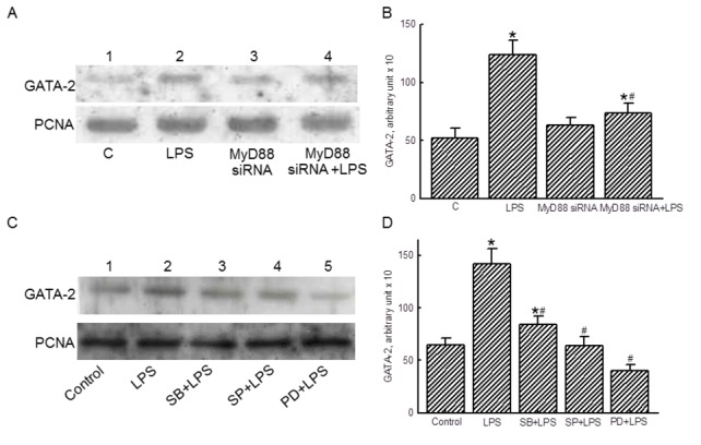
Figure 6. Effects of MyD88 small interference (si) RNA and MAPK inhibitors on translocation of GATA-2.

RAW 264.7 cells were exposed to lipopolysaccharide (LPS), MyD88 siRNA, and a combination of MyD88 siRNA and LPS. Amounts of GATA-2 were immunodetected (A, top panel). PCNA was measured as the internal control (bottom panel). These protein bands were quantified and statistically analyzed (B). RAW 264.7 cells were pretreated with 10 µM MAPK inhibitors, including SB203580 (SB), SP600125 (SP), and PD98059 (PD), for 1 h and then exposed to LPS. Nuclear GATA-2 was immunodetected (C, top panel). Amounts of PCNA were measured as the internal control (bottom panel). These protein bands were quantified and statistically analyzed (D). The immunoblotting results shown are a representative of 6 experiments, and the other statistically analyzed results are a compilation of 6 replications. Each value represents the mean ± SD. An asterisk (*) and pound sign (#) respectively indicate that the value significantly (p < 0.05) differed from the respective control and LPS-treated group.
