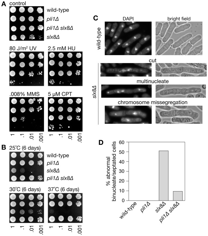
Figure 1. Cell growth, genotoxin sensitivity and chromosome segregation in slx8 Δ and pli1Δ single and double mutants.
(A) Spot assay comparing the genotoxin sensitivities of strains MCW1221, MCW4568, MCW4688 and FO986. Plates were photographed after 6 days growth at 30°C. (B) Spot assay comparing the growth at different temperatures of the same strains as in A. Plates were photographed after 6 days growth at the indicated temperature. (C) Example images of wild-type and slx8Δ binucleate and septated cells. (D) Percentage of binucleate and septated cells exhibiting abnormal chromosome segregation. A total of 100 binucleate/septated cells from two independent cultures were analysed for each strain.

