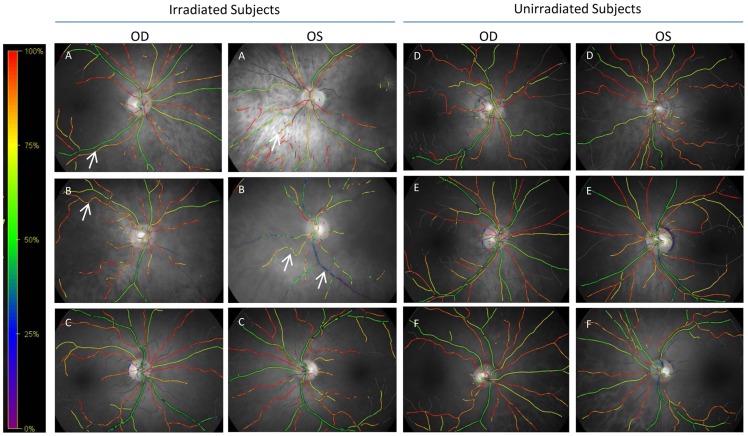
Figure 1. Representative qualitative retinal oximetry images of retinas from 3 irradiated (A–C) and 3 unirradiated (D–F) subjects from the two cohorts.
OD: Right eye. OS: Left eye. A color gradient with corresponding SO2 values is provided at the left of the images. The green/yellow vessels are venules while the red/orange vessels are arterioles. Some of the irradiated subjects exhibited highly variable SO2 values in venules (see arrow to blue venule in patient B, OS) or arterioles (see arrows in patient B, OD and patient A, OD and OS). Not all irradiated subjects exhibited this marked variability in qualitative SO2 measurement (Subject C). Unirradiated subjects had less within-subject variability (note the more consistent green venules and red arterioles in patients D–F).

