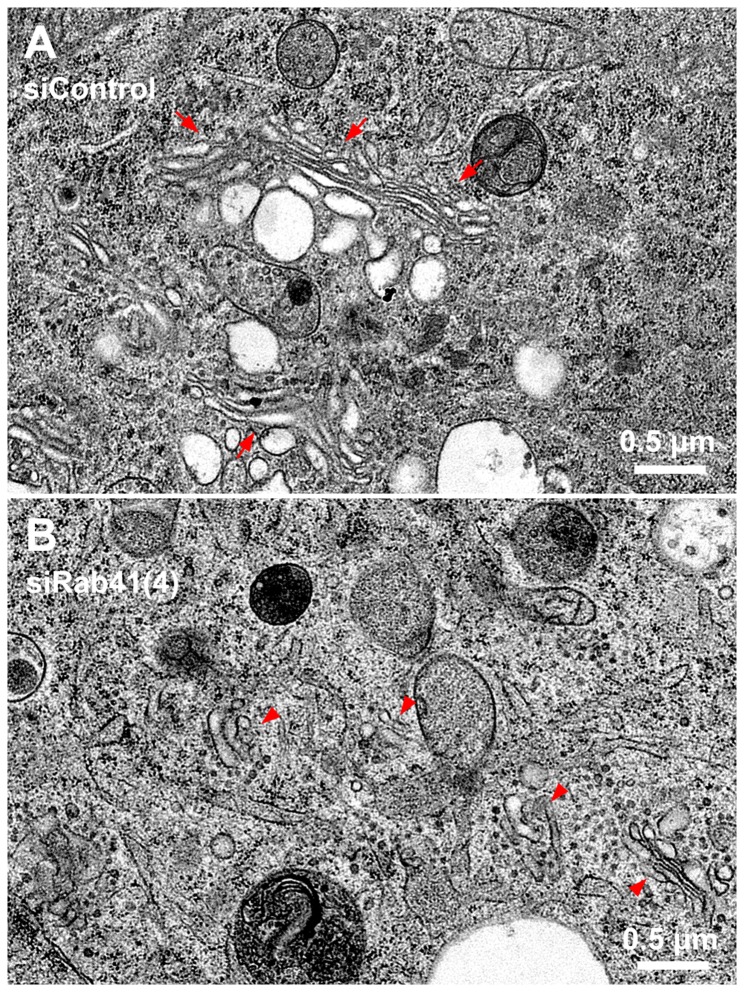
Figure 5. Electron microscopy demonstrates that Rab41 depletion causes dramatic Golgi fragmentation and accumulation of Golgi-associated vesicles.
HeLa cells stably expressing GalNAcT2-GFP grown on sapphire discs were treated with either control siRNA or siRab41(4) duplexes at a concentration of 200 nM. Cells were high-pressure frozen followed by freeze-substitution 96 h post initial transfection. Thin sections (50 nm) were collected to test the Golgi phenotype. In cells transfected with control siRNA, Golgi stacks were close enough to form a long Golgi ribbon-like structure (A, arrows). However, in cells treated with siRab41(4), short, isolated Golgi stacks (B, arrowheads) rather than Golgi ribbon were observed, and Golgi-associated vesicles accumulated (B).

