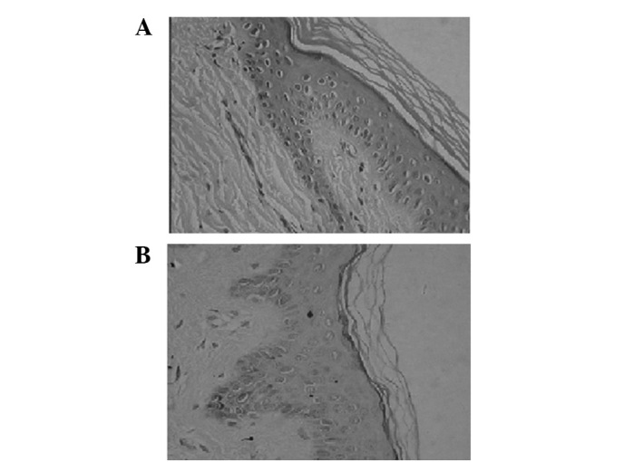
Figure 2.

(A) Two weeks after cografting, thick desquamation, active acanthocyte proliferation and an increased number of inflamed cells and fibroblasts between the dermal template and the raw surface juncture were observed (B) 12 weeks after cografting, the cograft skin mixed together well, the basal membrane was clear and continuous, the stratum corneum was mature, rete pegs extended downward, dermal collagen fibers had a uniform structure and there were fewer blood capillaries; however, there were no appendages of the skin. Hematoxylin and eosin staining; magnification, ×100.
