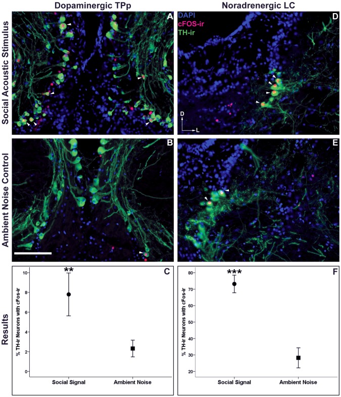
Figure 5. cFos-ir colocalization with catecholaminergic (TH-ir) neurons.
Arrowheads indicate cFos-ir colocalized to catecholaminergic neurons within the dopaminergic periventricular posterior tuberculum (TPp) (A, B) and the noradrenergic locus coeruleus (LC) (D,E) of males exposed to social acoustic signals and males exposed to ambient noise. Data in C and F are represented as mean percent colocalization ± SE, **p≤0.01, ***p<0.001. Scale bar = 100 µm. Arrows represent the dorsal (D) and lateral (L) orientation for each image.

