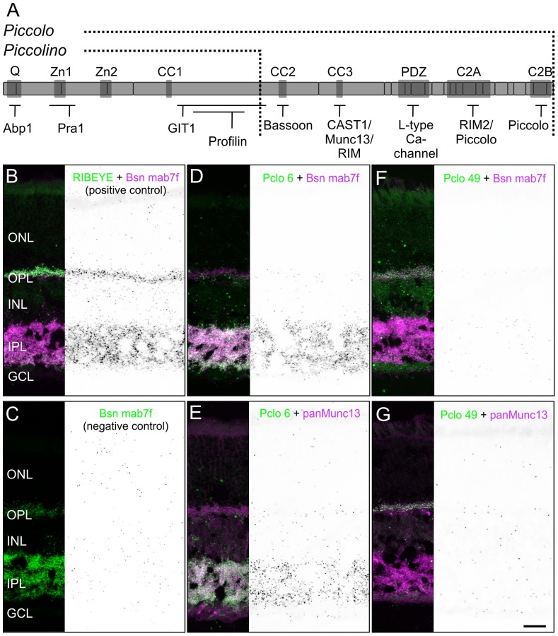
Figure 7. Missing interactions of Piccolino with Bsn and Munc13.
A: Schematic representation of full-length Pclo with its interaction domains (dark gray boxes) and known binding partners. The C-terminally truncated Piccolino lacks the C-terminal interactions. B–G: In situ proximity ligation assays (PLA) on vertical sections through wild-type retina (black and white panels) with corresponding fluorescence stainings. Positive control: interaction of RIBEYE and Bsn with the antibodies RIBEYE (green) and Bsn mab7f (magenta; B). Negative control: antibody Bsn mab7f (green) alone (C). Interaction of full-length Pclo with Bsn (D) and Munc13 (E) probed with the antibodies Pclo 6 (green), Bsn mab7f (magenta), and panMunc13 (magenta). Interaction of Piccolino with Bsn (F) and Munc13 (G) probed with the antibodies Pclo 49 (green), Bsn mab7f (magenta), and panMunc13 (magenta). ONL: outer nuclear layer; OPL: outer plexiform layer; INL: inner nuclear layer; IPL: inner plexiform layer; GCL: ganglion cell layer. Scale bar: 20 µm.

