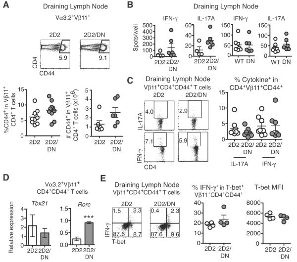
Figure 2. Notch inhibition in myelin-reactive CD4+ T cells does not alter initial activation or effector T cell differentiation.
(A) Percent and absolute number of Vβ11+CD4+CD44+ T cells in the draining lymph node (DLN) at peak disease (n=3–4 mice/group; ≥2 experiments); (B) Number of IFNγ and IL-17A-secreting cells as assessed by ELISpot in DLN from immunized WT, DN, 2D2, and 2D2/DN (n=3–4 mice/group; ≥2 experiments); (C) Frequency of IFNγ and IL-17A-producing DLN Vβ11+CD4+CD44+ T cells after restimulation with anti-CD3/CD28 and staining for intracellular cytokines (n=3–4 mice/group; 2 experiments); (D) Tbx21 and Rorc mRNA in activated CD44+ 2D2/DN and 2D2 Vα3+Vβ11+CD4+ T cells (n=3–4 mice/group; 2 experiments); (E) Intracellular T-bet and IFNγ in Vβ11+CD4+CD44+ T cells as assessed by intracellular flow cytometry (2 experiments; n=3–4 mice/group). Representative flow cytometry plots are shown. MFI: mean fluorescence intensity. p<0.001***.

