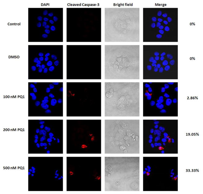
Fig. 4. PQ1 activates caspase-3 in T47D breast cancer cells.
T47D cells were treated with DMSO and various concentrations of PQ1 for 48 hours. Cells without treatments were used as controls. Expression of cleaved caspase-3 was determined by confocal microscopic analysis using immunofluorescence staining. Red indicates cleaved caspase-3 and blue indicates nuclei stained by DAPI. Percentages of cells with positive staining were labeled on the right of images.

