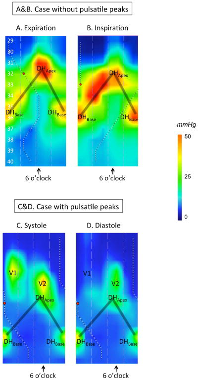
Figure 3.
Representative examples of 3D-HRM still images at expiration (A) and inspiration (B), illustrating the discrete pressure peaks within the EGJ. Figures 3C & 3D illustrate the case of a different subject with pulsatile peaks. The shaded ‘V’ on the topography plots indicates the disposition of the split diaphragm signal. See text for details of peaks DHApex, DHBase, V1, and V2.

