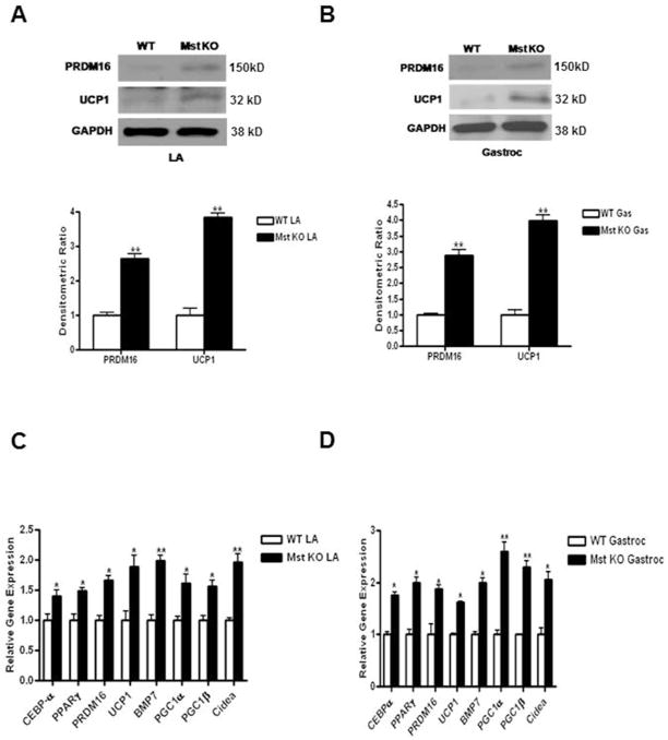
Fig. 1.
Analysis of PRDM16 and UCP1 protein expression in LA and Gastroc muscles isolated from WT and Mst KO mice. 100μg of total protein lysates from LA (A) and Gastroc (B) muscles were analyzed by Western blot (top panel). Quantitative densitometric analysis normalized to GAPDH is shown at the bottom panel. Analysis of a panel of BAT-specific mRNAs isolated from LA (C) and Gastroc (D) muscles of WT and Mst KO mice by quantitative real-time PCR. Experiment was repeated three times and representative data is shown (*, denotes p≤0.05; and **, denotes p≤0.005).

