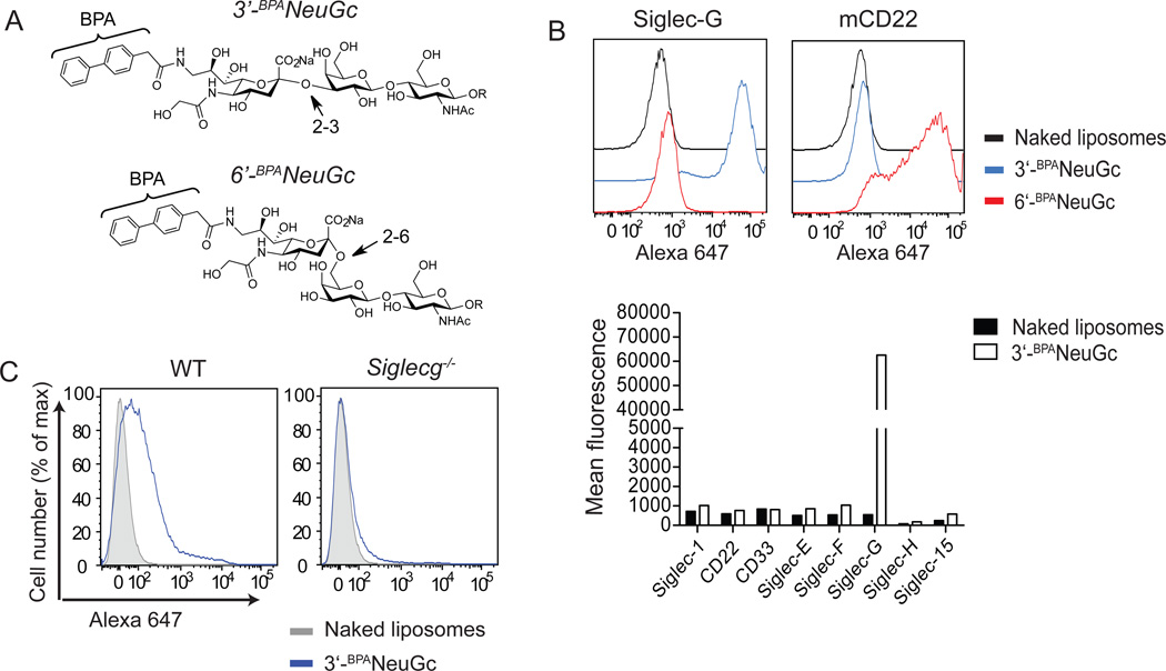
Figure 2. Development of a high affinity glycan ligand specific for Siglec-G.
A, Chemical structure of high affinity glycan ligands for Siglec-G (3’-BPANeuGc) and CD22 (6’-BPANeuGc). B, Top: Liposomes displaying the ligands for Siglec-G and CD22 bind specifically to cultured cells expressing Siglec-G or CD22, respectively. The cells were incubated with Alexa 647-labeled naked liposomes (black line), 3’-BPANeuGc liposomes (blue line), and 6’-BPANeuGc liposomes (red line), washed, and analyzed by FACS. Bottom: Liposomes displaying 3’-BPANeuGc do not bind to cells expressing any other murine siglec except Siglec-G. Binding of liposomes is expressed as mean channel fluorescence (MCF). C, Liposomes displaying the Siglec-G ligand bind to WT but not Siglec-G deficient B cells.

