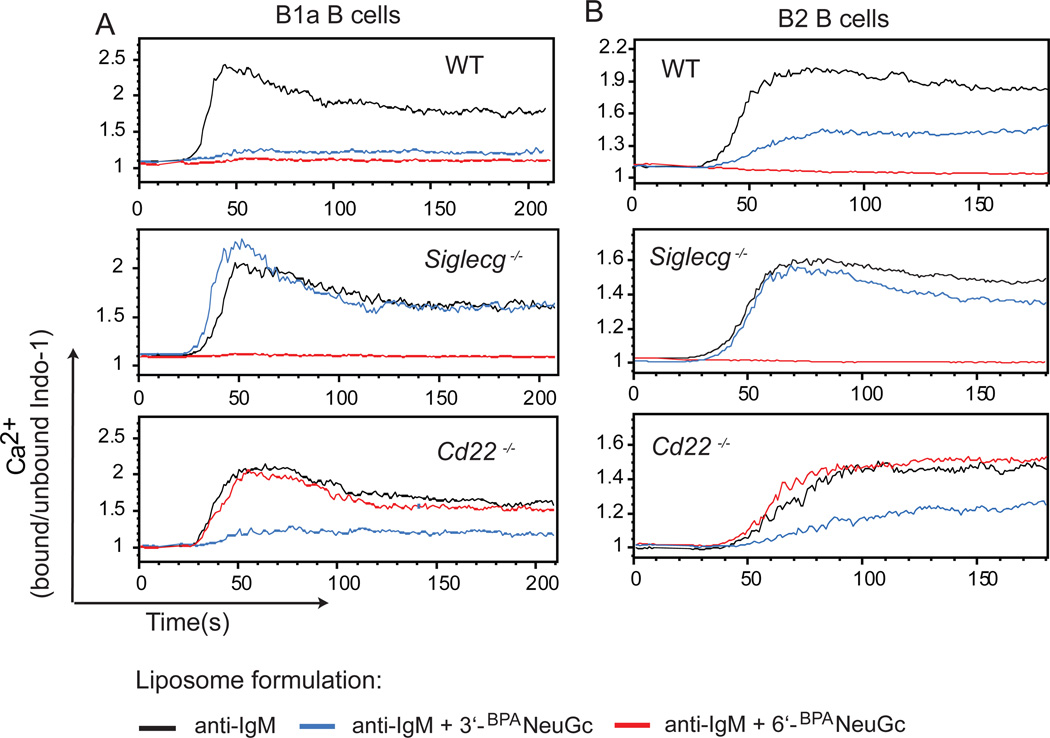
Figure 3. Inhibition of Ca2+-flux in B cells by STALs displaying 3’-BPANeuGc.
(A,B) Calcium flux of peritoneal cavity B1a (B220lowCD5+) B cells (A) or splenic (B220+CD5−) B2 B cells (B) from WT, Siglecg−/− or Cd22−/− mice. Cells were stimulated at t=10 s with liposomes displaying anti-IgM (black), anti-IgM + 3’-BPANeuGc (blue), or anti-IgM + 6’-BPANeuGc (red) and the intracellular Ca2+-mobilization was measured by FACS.

