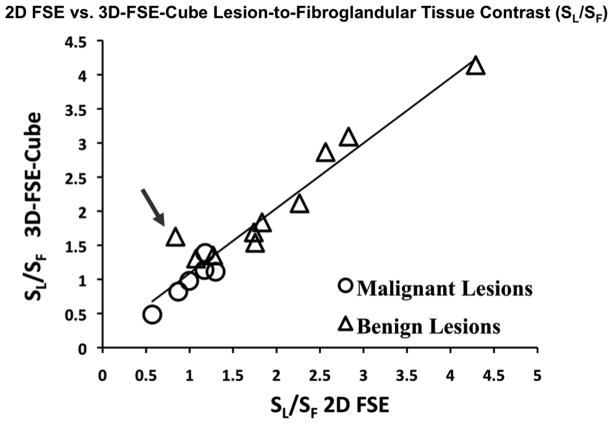
Figure 2.
Lesion-to-fibroglandular tissue signal ratio (SL/SF) is highly correlated between 2D FSE and 3D-FSE-Cube (R2 = 0.93). The data point indicated by the arrow was measured in a very small fibroadenoma (images shown in Figure 5) in which partial voluming may have diminished the signal level in the lesion on 2D FSE, demonstrating a case in which the increased resolution available with 3D-FSE-Cube allows for a more accurate depiction of lesion signal intensity.

