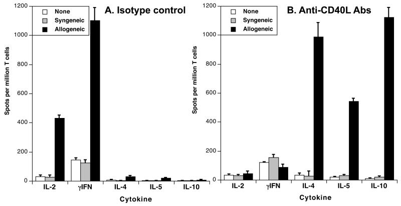
Figure 6. MR1 treatment promotes the activation of T cells secreting type 2 cytokines.
C3H mice were transplanted with a B6 conventional skin allograft and injected with medium (panel A) or MR1 anti-CD40L mAbs as described earlier. Ten days later, spleen T cells were collected and restimulated in vitro with PBS (white bars), syngeneic APCs (grey bars) or irradiated donor APCs (solid bars) (direct allorecognition). The frequencies of activated T cells secreting type 1 (IL-2 and γIFN) and type 2 (IL-4, IL-5 and IL-10) cytokines were measured by ELISPOT. The results are expressed as numbers of cytokine-forming spots per million T cells ± SD and are representative of 3-5 mice tested individually in each group.

