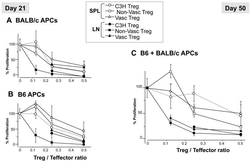
Figure 7. Treg suppression assays.
Naïve CD4+CD25− T effector cells from graft recipient-type (C3H) were incubated with CD90-depleted irradiated splenocytes from donor B6, third party (BALB/c), or a 1:1 mixture of BALB/c and B6 cells. CD4+CD25+ Treg cells were isolated from naïve C3H mice (circles) and C3H mice transplanted with either a conventional (squares) or a vascularized (triangles) B6 skin allograft. Tregs were added to the coculture at the indicated Treg/T effector ratios (X-axis), and suppression of alloreactive cell proliferation was measured by thymidine incorporation. (A and B): suppression of alloresponses to third-party (A) or donor (B) by spleen (SPL, opened symbols) and lymph node (LN, closed symbols) Tregs collected 21 days after transplantation. (C): Similar studies were performed using Tregs collected at d50 post-transplantation from spleen and lymph nodes and CD4+ T effector cells stimulated by an equal number of B6 and BALB/c APCs.

