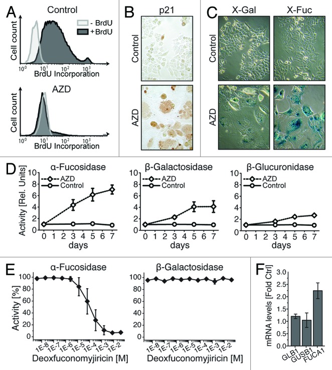
Figure 1. α-Fucosidase is a sensitive biomarker of cellular senescence. HCT116 cells were induced to undergo senescence by 7 d of treatment with the Aurora inhibitor AZD1152-HQPA (AZD; 500 ng/mL). (A) FACS analysis reveals BrdU uptake in control but not drug-treated cells. (B) Induction of p21 expression upon AZD treatment. (C) Parallel cytochemical analyses show a more intensive blue staining for α-Fuc as compared with β-Gal using X-Fuc and X-Gal substrate. (D) Fluorometric detection of β-Gal, α-Fuc and β-Gluc activities after indicated times of AZD treatment. One unit equates to the activity in control samples. (E) Protein lysates from cells treated for 7 d with AZD were analyzed for α-Fuc and β-Gal activities in the presence of the α-Fuc inhibitor deoxyfuconojirimycin. (F) qRT-PCR analysis of GLB1, GUSB1 and FUCA1 mRNA after AZD treatment. Panels (A–C) show representative data and panels (D–F) mean values ± SD, each from three independent experiments.

An official website of the United States government
Here's how you know
Official websites use .gov
A
.gov website belongs to an official
government organization in the United States.
Secure .gov websites use HTTPS
A lock (
) or https:// means you've safely
connected to the .gov website. Share sensitive
information only on official, secure websites.
