
Figure 3.
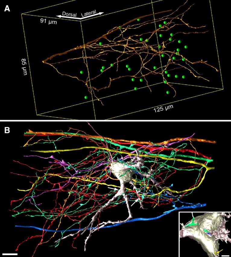
Exuberant innervation of MNTB at P2. A, Reconstruction of single large-diameter axon oriented mediolaterally in image volume. Green spheres indicate center of MNTB cells (35 of 71 cells in volume) with synaptic contact from this axon onto soma or dendrites. Yellow lines depict outline of image volume. B, To illustrate convergence of multiple inputs, a cell contacted by five large-diameter trapezoid body axons on dendrites (white arrows; left arrow indicates contact on opposite side of dendrite) or cell body [inset, green, blue, and yellow (arrow highlights ASA); fourth ASA not shown] is shown. Contacts occurred via branches from parent axon. Scale bars: B, 10 μm; B, inset, 2.5 μm.