
Figure 7.
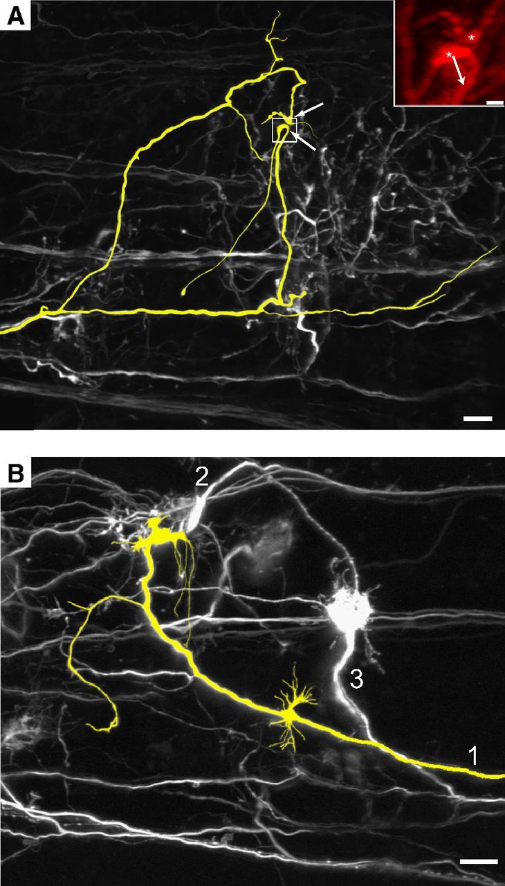
Branches of parent axon do not converge onto the same MNTB cell. A, Subset of axons electroporated with Alexa Fluor 488 dye and imaged using two-photon microscopy in P3 animal. Images in both panels are of the collapsed image stack. Only example of axon and branches that terminate near each other (colorized yellow; white arrows indicate terminals). Inset, Close-up of boxed region viewed at angle to demonstrate curvature of large ending (arrow) away from smaller ending (both indicated with asterisks), likely indicating innervation of different postsynaptic structures. B, Typical pattern of branching and innervation where axon did not branch at medial locations (area not shown to right of B) and branches terminate in distinct regions in the MNTB. Axons 1 and 2 terminate via large endings near each other (P4 animal). Scale bars: A, B, 20 μm; A, inset, 5 μm.