Figure 3.
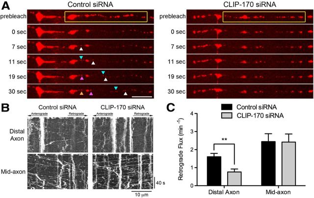
CLIP-170 is necessary for efficient transport initiation from the distal axon. A, Time series of LAMP1-RFP motility in the distal axon of DRG neurons imaged at 4 DIV. DRG neurons were transfected with either control siRNAs or siRNA against CLIP-170. The yellow box demarcates the photobleached zone; 0 s is the first frame after photobleaching. The different colored arrowheads demarcate LAMP1-RFP-positive cargos moving into the photobleached zone. Significantly fewer cargos entered the photobleached zone after CLIP-170 knock-down. Scale bar, 10 μm. B, Kymographs of the time series of the distal axon LAMP1-RFP motility shown in A and the mid-axon LAMP1-RFP motility. Scale bars, 10 μm and 40 s for the x and y axes, respectively. C, Quantification of distal and mid-axon retrograde flux after photobleaching. Flux was determined by counting the number of retrograde vesicles that moved >3.5 μm into the photobleached zone. Data are shown as mean ± SEM; for distal and mid-axon flux, n = 19–20 neurites per condition from two independent experiments; **p < 0.01, Student's t test.

