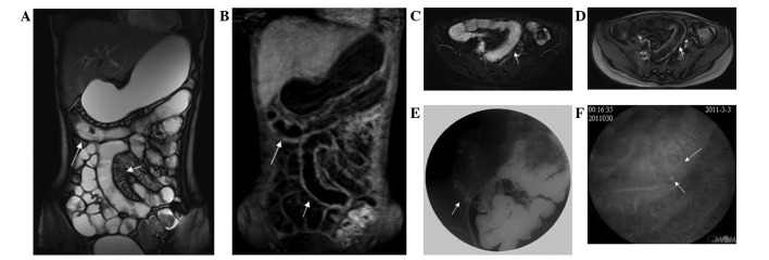
Figure 2.

Correlation between MRE, CGR and VCE in a 16-year-old female patient with Crohn’s disease confirmed at histology. (A) Coronal T2-weighted TrueFISP image revealed that a dilated ileal loop with irregular wall thickening, increased mesenterial vascularity, separation of loops (white short arrow) and transverse colon wall thickening (white long arrow); (B) enhanced coronal T1-weighted image revealed wall thickening of the ileum (white short arrows) and transverse colon (white long arrows) with contrast enhancement; (C) axial T2-weighted fat suppression images revealed diffuse thickening and edema of the ileum (white short arrow); (D) enhanced axial T1-weighted image revealed diffuse thickening and edema of the ileum (white short arrow); (E) the CGR image revealed luminal stenosis of the distal ileum (short white arrows), mucosal fold thickening, edema and broadening of the fat space around the intestine; (F) VCE clearly revealed distal ileum mucosal changes with ulcerations (short white arrow), congestion and edema (long white arrow). MRE, magnetic resonance enterography; CGR, conventional gastrointestinal radiography; VCE, video capsule endoscopy; TrueFISP, true fast imaging with steady-state precession.
