
Figure 1.
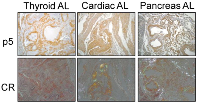
Histochemical analysis of p5 binding to AL amyloid deposits in tissue sections. Biotinylated p5 was added to AL-containing tissue sections (p5) and binding evidenced by brown diaminobenzidine staining. Co-localization with amyloid deposits was shown by staining consecutive tissue sections with Congo red (CR) and examining the presence red-green birefringence indicative of amyloid.