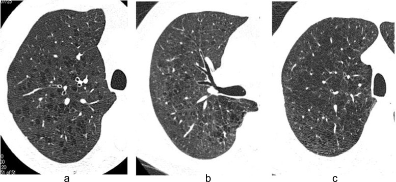
Fig. (1).
HRCT images of three subtypes. Typical high-resolution CT images in Subtype A (a), Subtype B (b), and Subtype C (c) are shown. Subtype A is defined as emphysema which showed round or oval shape LAA with well-defined border. Subtype B is defined as emphysema which showed polygonal or irregular-shaped LAA with ill-defined border. Subtype C is defined as emphysema which showed irregular-shaped LAA with ill-defined border coalesced with each other.

