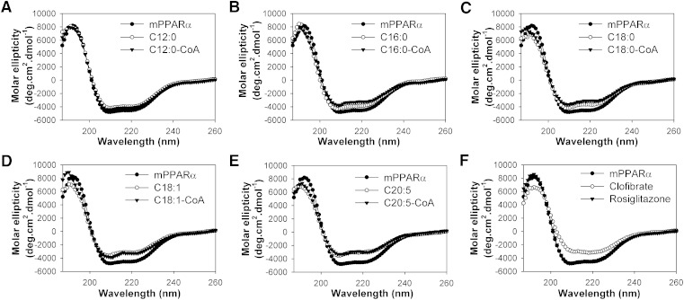
Fig. 6.
Far UV CD spectra of mPPARα in the absence (filled circles) and presence of added ligand: (A) lauric acid (open circles) or lauryl-CoA (filled triangles); (B) palmitic acid (open circles) or palmitoyl-CoA (filled triangles); (C) stearic acid (open circles) or stearoyl-CoA (filled triangles); (D) oleic acid (open circles) or oleoyl-CoA (filled triangles); (E) EPA (open circles) or EPA-CoA (filled triangles); and (F) clofibrate (open circles) or rosiglitzone (filled triangles). Each spectrum represents an average of 10 scans for a given representative spectrum from at least three replicates.

