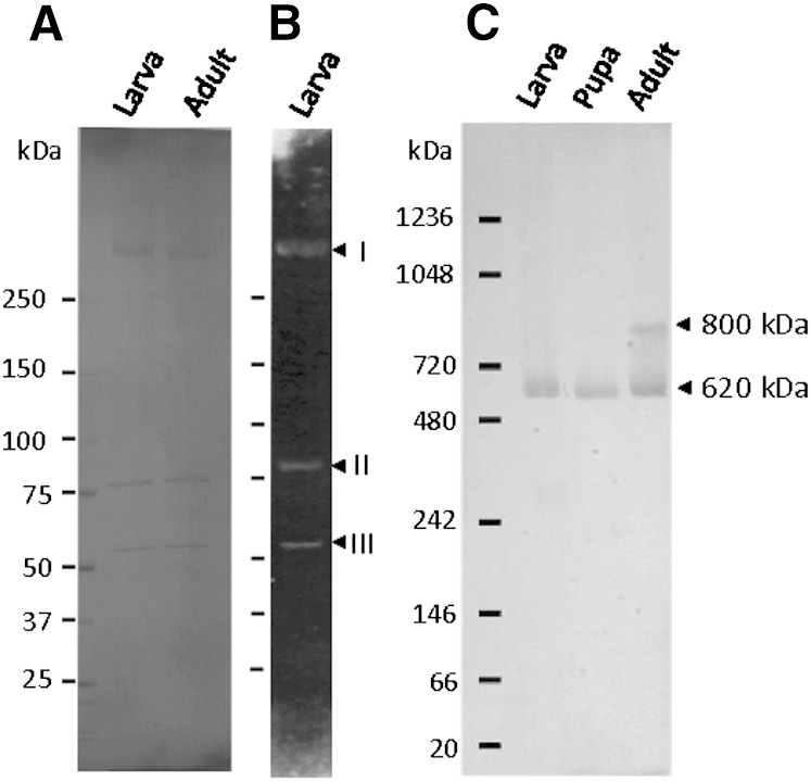
Fig. 6.
Estimation of the molecular mass of native LTP. A: Purified LTP from the hemolymph of fifth instar larvae and adults was subjected to SDS-PAGE and stained with Coomassie Brilliant Blue R-250. B: Purified LTP from the hemolymph of fifth instar larvae was subjected to SDS-PAGE and transferred to a PVDF membrane. The blot was incubated with FITC-Con A solution. Detection of glycoproteins bound to FITC-Con A was carried out under ultraviolet light. Arrows indicate apoLTP-I (I), apoLTP-II (II), and apoLTP-III (III) from the top, respectively. C: One microliter each of the hemolymph from fifth instar larva, pupae, and adults was electrophoresed by 3–10% blue-native PAGE. Separated proteins were transferred to a PVDF membrane. Native LTP was detected by Western blot analysis using the anti-apoLTP-I antibody. Arrows indicate the 620 kDa and 800 kDa LTP. Numbers on the left of each panel represent the molecular masses for protein standard.

