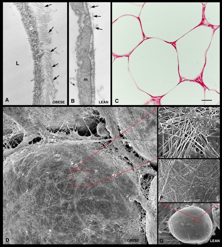
Fig. 4.
Extracellular matrix of visceral adipose tissue from genetically obese mice. TEM shows numerous collagen fibrils (arrows) in close contact with the external surface of a hypertrophic adipocyte in genetically obese mice (A) and a small number of collagen fibrils on the external surface of the adipocyte basal lamina in control mice (B). By light microscopy (C), strong picrosirius red staining confirms the presence of large amounts of collagen in the extracellular matrix surrounding the hypertrophic adipocytes. HR-SEM shows an intricate network of collagen fibrils covering the surface of a hypertrophic adipocyte (D). In (G), a control adipocyte is shown for comparison. (E and F) Enlargements of the areas framed in (D) and (G), respectively. L, lipid droplet; m, mitochondria. Scale bar: 0.3 μm for (A) and (B); 30 μm for (C); 5 μm for (D); 1 μm for (E); 0.7 μm for (F); 6 μm for (G).

