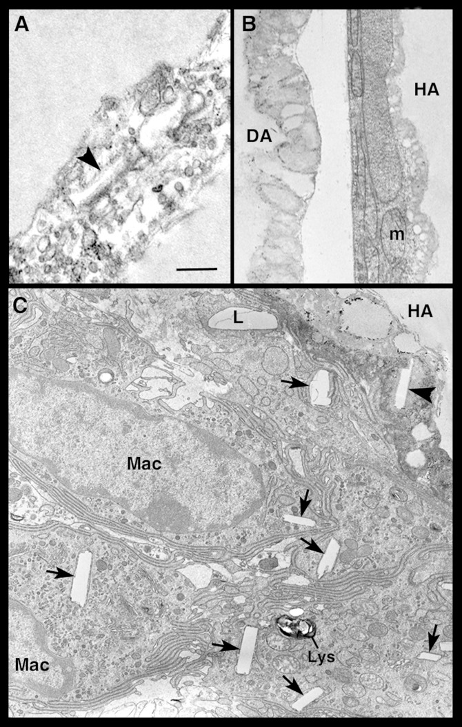
Fig. 6.
Transmission electron microscopic images of hypertrophic adipocytes in visceral fat of db/db mice. In (A), the cytoplasm of two closely apposed hypertrophic-degenerating adipocytes is disrupted, devoid of recognizable organelles, and contains vacuoles and a cholesterol crystal (arrowhead). In (B), a hypertrophic adipocyte (HA) with well-preserved organelles is found near a degenerating adipocyte (DA), whose cytoplasm lacks organelles, which are replaced by lipid-like material. In (C), several macrophages (Mac) are closely apposed to a HA containing a cholesterol crystal (arrowhead) in the degenerating cytoplasm; the macrophages contain lipid droplets (L) and numerous cholesterol crystals (arrows). m, mitochondria; lys, lysosome. Scale bar: 0.6 μm for (A); 1 μm for (B) and (C).

