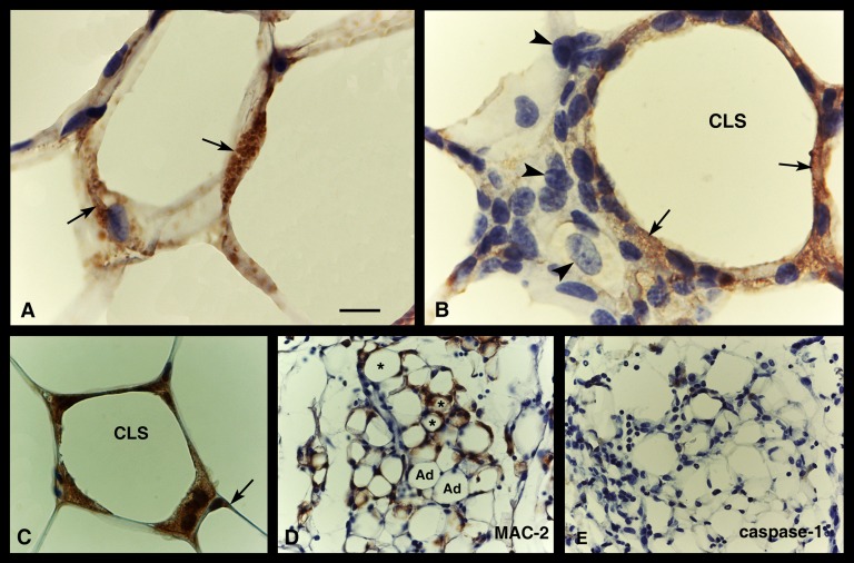
Fig. 9.
NLRP3 activation in adipose tissue of genetically obese mice. Immunohistochemistry. Caspase-1 immunostaining is evident (brownish precipitate, arrows) in hypertrophic (or degenerating) adipocytes (A) and in a CLS (B) of the visceral fat of a db/db mice: here, staining is found in macrophages surrounding the large lipid droplet but not in those lying at a distance from it (arrowheads), which are not yet engaged in lipid clearing. In (C), ASC immunoreactivity is found in a CLS, but weak staining is also visible in the cytoplasmic rim of a hypertrophic adipocyte (arrow). In (D) and (E), serial sections of visceral adipose tissue of FAT-ATTAC mice are stained with MAC-2 and caspase-1, respectively: MAC-2-positive CLSs (asterisks) and apoptotic adipocytes (Ad) are both negative for caspase-1. Scale bar: 11 μm for (A) and (B); 18 μm for (C); 22 μm for (D) and (E).

