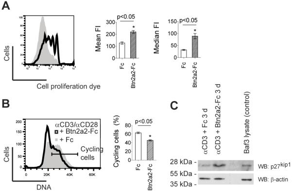
Figure 2. Btn2a2 inhibited entry into the cell cycle of anti-CD3 and anti-CD28-activated CD3+ primary T cells.

T cells from spleen and lymph nodes were stimulated for 4 d using 1 μg/ml anti-CD3, 1 μg/ml anti-CD28, and 10 μg/ml fusion protein. (A) Cell proliferation of cells 4 d after activation in Btn2A2-Fc (black outline) or Fc control (grey area) visualized with 1 μM eFluor670. (B) DNA content visualized using propidium iodide. % cycling cells (horizontal bar) calculated by subtracting the two-fold percentage of cells in the left hand part of the G0/G1 peak from 100%. (C) 4 d after activation of cells with 1 μg/ml anti-CD3 in presence of Fc control or BTN2A2-Fc, expression of the cell cycle entry inhibitor p27kip1 was analysed by western blotting for p27kip1. 1×10⁵ cells per lane. Mouse Baf/3 cells were positive control for p27kip1. Loading control: β-actin. FACS data representative of 3 experiments with 3 replicates. Statistically significance as for Fig. 1.
