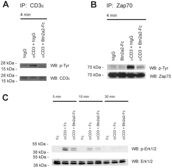
Figure 3. Btn2a2 co-ligation inhibits T cell receptor signaling in anti-CD3 activated T cells.

(A) 2B4 cells were stimulated for 4 min with 1 μg/ml anti-CD3 (clone 2c11) in 10 μg/ml Btn2a2-Fc or hIgG. Control: 2B4 cells stimulated with 10 μg/ml hIgG. After immunoprecipition with anti-CD3ε (clone CD3-12) and SDS-PAGE analysis was for tyrosine phosphorylation (4G10). (B) 2B4 cells stimulated 5 min with 1 μg/ml anti-CD3 in 10 μg/ml Btn2a2-Fc or hIgG. After lysis, samples were immunoprecipitated with biotinylated anti-Zap70 antibody, treated as in A and analysed for phosphorylation (4G10). (C) CD3+ primary T cells were stimulated with 1 μg/ml anti-CD3 and 10 μg/ml fusion protein, treated as above, and analysed for phosphorylation of Erk1 and Erk2 (p-Erk1/2). Antibody to total Erk1/2 (Erk1/2) was used for loading. Data from >2 independent experiments.
